Tissue support
When the residual ridge is capable of supporting the denture and withstanding functional stresses, it facilitates even load distribution across the supporting tissues, thereby enhancing oral health outcomes.
Several key factors are taken into account when assessing the adequacy of tissue support, including:
- The length of the residual ridge
- The contour of the residual ridge
- The quality and quantity of the supporting bone
- The characteristics of the overlying mucosa
The length of the residual ridge
The length of the edentulous space plays a critical role in determining the extent of tissue support available for the denture. As the edentulous space increases, the tissue-borne support for the denture base also becomes more significant (Figure 1-23). The region adjacent to the abutment tooth derives its primary support from that tooth, whereas areas further from the abutment rely predominantly on the residual ridge for support in distal-extension RPDs.
The contour of the residual ridge
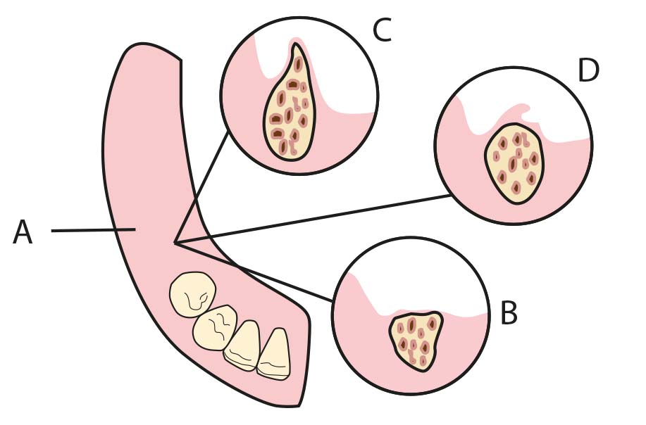
The contour of the residual ridges also plays an important role in distributing the forces generated by the function of the partial denture (Figure 1-23). Broad, well-formed ridges absorb more stress than thin, small, knife-edge ridges. Parallel-sided, wide ridges enable the fabrication of a denture base with extended flanges, which contribute to enhanced stability by resisting lateral displacing forces.
The quality and quantity of the supporting bone
The efficiency of support is largely dependent on the structural quality of the remaining ridge underneath the prosthesis. In particular, areas with reduced cortical bone density exhibit a diminished capacity to withstand functional stresses. Radiographic evaluation serves as a valuable tool in assessing the bone’s response to occlusal loading and the dynamic movements of a distal-extension denture base. These assessments also aid in predicting the bone’s ability to function as a reliable support for both existing and future prosthetic appliances.
The characteristics of the overlying mucosa
Both the thickness of the mucosa over the ridge and the total surface area covered by the denture base influence the magnitude and distribution of functional forces. The mucosa covering the residual ridge plays a significant role in providing support. While some regions of the mucoperiosteum are firm and immobile, others consist of mobile and resilient tissue. Soft, thin, and displaceable tissues offer minimal support to the prosthesis and contribute little to the lateral stabilization of the denture base (Figure 1-23).
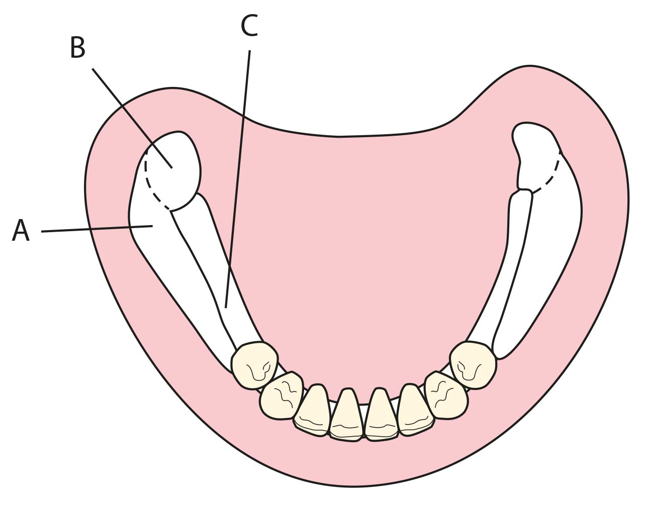
Supporting tissue areas in the mandible (Figure 1-24):
• Buccal shelf area (primary support),
• Retromolar pad area (primary or secondary support depending on tissue density),
• Slopes of the residual crest (secondary support).
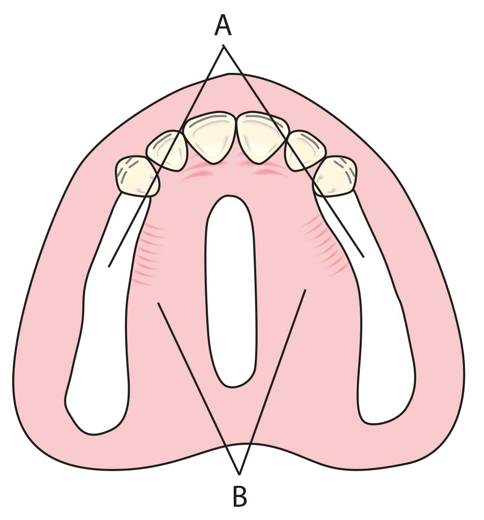
Supporting tissue areas in the maxilla (Figure 1-25):
• Crest of the posterior residual crest (primary support),
• Slopes of the residual crest (secondary support),
• Horizontal parts of the hard palate (primary support).