
Oral surgical preparation
Pre-prosthetic surgical procedures should be completed as early as possible. Periodontal, surgical, and endodontic treatments are planned in a way that they conclude simultaneously and allow sufficient time for healing. The healing period between surgical and restorative procedures should be at least 6 weeks, preferably 3 to 6 months.
Extractions
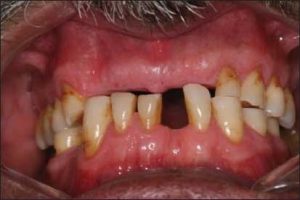
Following diagnostic cast surveying and treatment planning, tooth extractions should be carried out early enough to allow adequate healing. However, each tooth should be evaluated based on its location and potential contribution to the success of the treatment. Teeth that are strategically important and that will support the success of the prosthesis should be retained, while those with a questionable prognosis or that may complicate treatment planning should be extracted (Figure 10-1).
Removal of residual roots
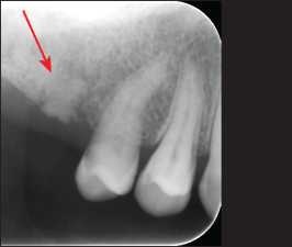
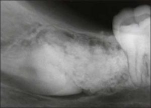
All retained roots or root fractures that are close to the tissue surface or exhibit pathological signs should be removed (Figure 10-2).
Impacted teeth
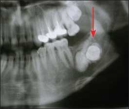
Impacted teeth located in edentulous areas and exhibiting pathological findings (Figure 10-3) should be removed, similar to retained roots. However, asymptomatic impacted teeth that are completely surrounded by bone may be retained—especially in older patients—in order to preserve arch morphology, provided that the patient is properly informed and the condition is monitored.
Malposed teeth
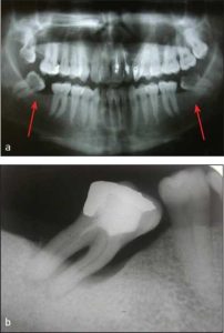
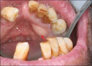
The loss of a single tooth or a group of teeth may lead to malposition of the remaining teeth due to their extrusion or tipping (Figure 10-4).
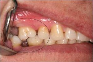
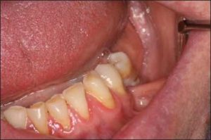
In most cases, the alveolar bone moves occlusally along with the extruded tooth (Figure 10-5). Although not always a practical solution, orthodontic treatment may be beneficial in managing occlusal discrepancies. Another treatment option is the surgical correction of the position of the tooth and the supporting alveolar bone.
Cysts and odontogenic tumors
Panoramic radiography is recommended for the evaluation of unexpected pathological conditions. If a suspicious finding is detected, periapical radiographs should be taken (Figure 10-6), and consultation should be requested when necessary.
Exostoses and tori
The mucosa over abnormal bony growths such as exostoses and tori, which may affect the planning of removable partial dentures, is thin and susceptible to trauma (Figure 10-7). Consequently, prosthetic components in contact with these areas can cause irritation and chronic ulceration. Prosthesis design should either avoid these regions or provide adequate relief. When neither option is feasible, surgical intervention is indicated.
Hyperplastic tissue
Hyperplastic tissues may present as fibrous tubercles, soft fibrous ridges, epulis, or papillary papillomatosis (Figure 10-8). Surgical treatment should be performed on these tissues to create a firm support for the prosthesis. This approach enhances prosthesis stability, reduces harmful forces on abutment teeth and tissues, and often results in a more suitable occlusal plane for tooth arrangement. During the postoperative healing period, the fabrication of a surgical stent can improve patient comfort and help maintain vestibular depth. The previous removable prosthesis can also be adjusted and used as a surgical stent.

Muscle attachments and frena
Due to the reduction in ridge height as a result of resorption, muscle attachments may be located on or near the residual ridge crest (Figure 10-9). The mylohyoid, buccinator, mentalis, and genioglossus muscles can contribute to such problems. Additionally, the mentalis and genioglossus muscles may cause bony prominences in their attachment areas, which can complicate prosthetic planning. Ridge augmentation procedures that relocate muscle attachments and remove bony prominences improve the comfort and function of removable dentures.
The maxillary labial and mandibular lingual frenula are the most common frenula causing difficulties in shaping the denture base. Frenula that extend to the ridge crest can be easily corrected surgically; otherwise, they may reduce denture retention and cause prosthesis fractures.
Knife-edge ridges and bony spines
Sharp bony spines (Figure 10-10) and knife-edge ridges should be smoothed with minimal bone removal. If smoothing results in a significant reduction of ridge height, ridge augmentation or vestibuloplasty procedures may be recommended.
Polyps, papillomas, and traumatic hemangiomas
All abnormal soft tissue lesions should be excised and subjected to pathological examination prior to the fabrication of a removable partial denture. Even if these lesions have not caused any prior complaints, trauma induced by the prosthesis may lead to malignant transformation of the soft tissue lesions.
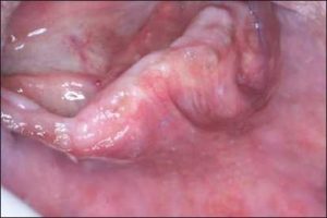
Hyperkeratoses, erythroplasia, and ulcerations
All abnormal white, red, or ulcerative lesions should be examined regardless of their association with the denture base or framework. If the lesion is larger than 2 mm, a biopsy with a margin of at least 5 mm should be performed for evaluation.